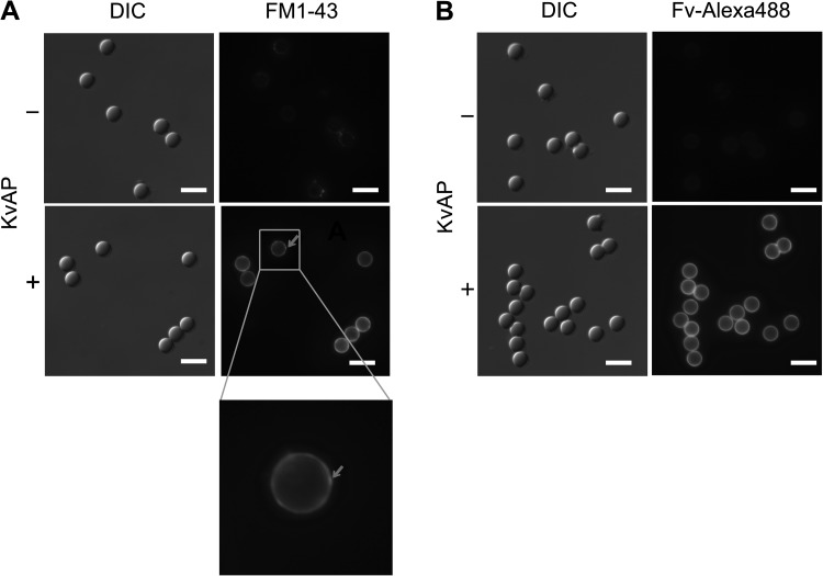
Figure 3.
Surface-anchored ion channels guide the formation of bSUMs. KvAP channels were used as a model system to form bSUMs around silica beads. The bSUMs around the beads of 5 µm in diameter were stained with FM1-43 (A) or with the KvAP-specific Fv–Alexa Fluor 488 (B). The fluorescence images (right) were shown side-by-side with the DIC images (left). Without KvAP, no fluorescence was seen around the beads (top rows). There was weak, nonspecific binding of FM1-43 to the beads. With KvAP, in bSUMs, there was uniform, circular staining around individual beads by FM1-43 and Fv–Alexa Fluor 488 (bottom right in both A and B). The integration of the epifluorescence signal made the edge of a vesicle much brighter than its center. The red arrow in A points to a small puncta, which is better seen in the zoomed-in view underneath and is quite likely caused by the attachment of a small vesicle. Bars, 10 µm.

