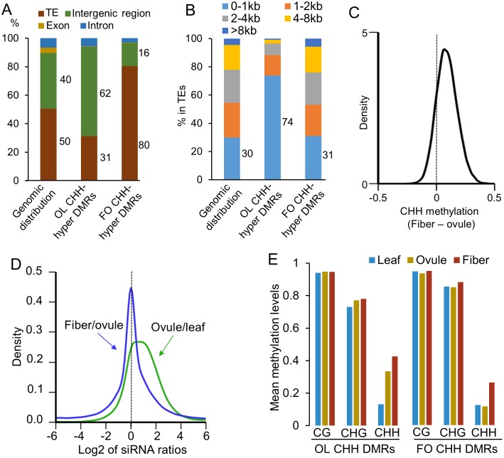
Fig 2. Genomic distribution of differentially methylated regions (DMRs).
(A) Percentage of DMRs in TE (brown), intergenic region (green), exon (dark yellow) and intron (blue). (B) Percentage of DMRs corresponding to TEs with different sizes. (C) Kernel density plot of CHH methylation change of fiber/ovule in OL CHH-hyper DMRs. (D) Kernel density plot showing 24-nt siRNA fold-changes of ovule/leaf in OL CHH-hyper DMRs (green) and of fiver/ovule in FO CHH-hyper DMRs (blue). (E) Percentage of CG, CHG and CHH methylation in the leaf (blue), ovule (yellow), and fiber (red) in OL CHH-hyper DMRs and FO CHH-hyper DMRs.

