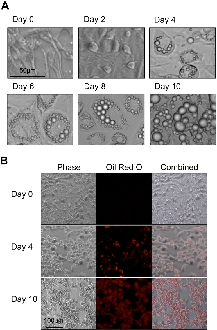
Fig 2. 3T3-L1 adipocyte differentiation.
(A) Phase contrast images of 3T3-L1 cells from induction (day 0) to ten days post-induction. Cells initially display a fibroblast phenotype. Over the course of differentiation, cell morphology changes and cells accumulate lipid droplets internally. (B) Triglyceride staining of 3T3-L1 cells with Oil Red O. Initially, the fibroblast phenotype displays negligible staining. As the adipocytes mature, the amount of staining increases until almost the entire cell volume is stained red.

