Fig. 4.
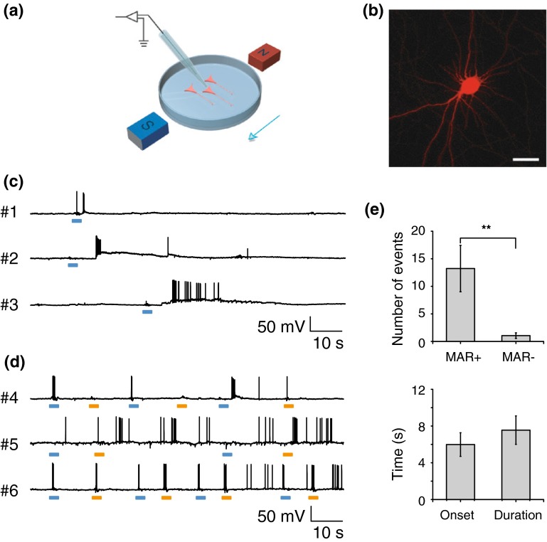
Neuronal spiking activity driven by the magnetic field via MAR. a Experiment scheme of whole-cell patch-clamp recording. Magnetic stimulation was achieved through a pair of handheld magnets. b Confocal imaging of a typical MAR-p2A-mCherry expressing neuron. Scale bar, 30 μm. c Current-clamp recording showing changes of membrane potential to magnetic stimulation. Three example neurons exhibited membrane depolarization and increasing firing rate to the onset of the magnetic field. Scale bar, 10 s, 50 mV. d MAR triggered action potentials displayed on-response and off-response firing patterns. Voltage traces of three representative neurons showed distinct firing patterns in response to magnetic field-on and field-off. Upper, the neuron only fired action potentials to the onset of magnetic field. To the opposite, the neuron shown in middle panel mainly responded to the removal of magnet. Another group showed typical firing pattern (lower panel) that both switch-on and switch-off of magnetic field elicited action potentials. Blue bar, field-on; orange bar, field-off. e Magnetic field induced significant increase in number of action potentials with mean onset latency of 5.3 ± 1.1 s and average duration of 8.5 ± 1.5 s when compared to spontaneous firing rate (13.2 ± 4.2 spikes versus 1.0 ± 0.5 spikes; n = 19; **, P < 0.01, paired t test). Error bar, s.e.m. Spikes were counted in 20 s after the first elicited spike within 20 s after the magnetic field was turned on

