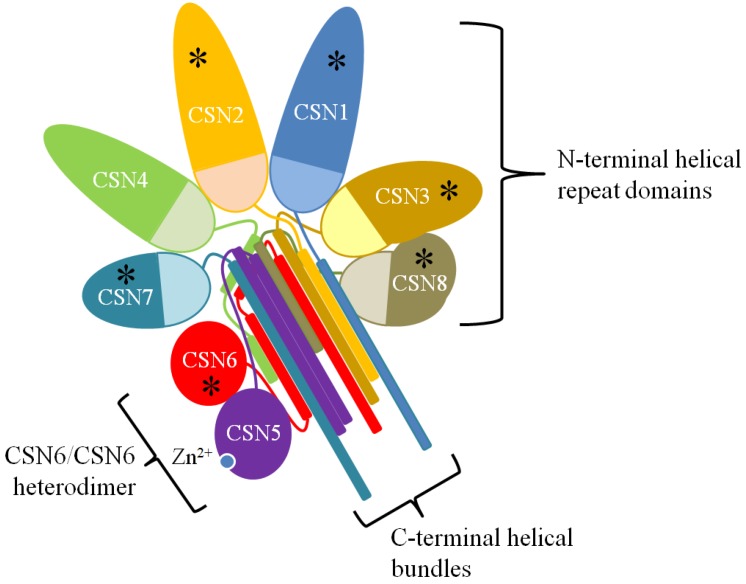
Figure 1.
The CSN Structure. A two-dimensional schematic representation of the three-dimensional structure of the CSN as determined by Lingaraju et al [34]. The N-terminal repeat domains radiate out from the winged-helix domains of the PCI ring (lightly shaded half-circles). The C-terminal helical regions form a helical bundle that stabilizes the complex. The MPN domains of CSN5 and CSN6 rest on the helical bundle. Subunits reported as phosphorylation targets are marked with an asterisk (*).

