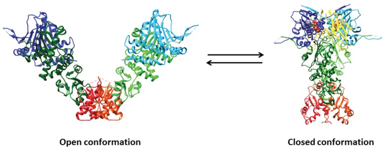
Figure 1.
Crystal structures of open (i.e., ligand-free) full-length Hsp90 from E. coli (HtpG; PDB ID: 2IOQ) and closed (i.e., ATP- and p23-bound) yeast Hsp90 (PDB ID: 2CG9). The N-domain is depicted in blue and cyan, the middle domain in dark green and light green, and the C-domain in red and orange. The p23 co-chaperone is in yellow, whereas ATP is depicted through a space-filling representation. Pictures were drawn by UCSF-Chimera [92].

