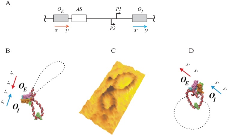
Figure 11.
Atomic force microscopy of GalR-HU mediated DNA loops. (A) Sketch of the gal DNA with a red arrow in the direction of 5' to 3' for OE and with a blue arrow in the direction of 5' to 3' for OI; (B) Model of antiparallel loop with the 3' of OE and OI facing each other in a head-to-head arrangement; (C) AFM results showing an antiparallel loop as predicted in model (B); (D) Model of parallel loop with the 3' of OE and OI facing opposite direction in a tail-to-head arrangement (reproduced with permission from Elsevier [72]).

