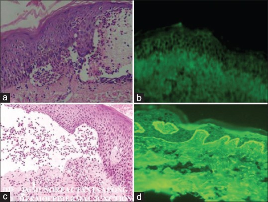
Figure 3.

(a) Hematoxylin and eosin of pemphigus vulgaris showing intraepidermal cleft [×400] and (b) direct immunoflourescence showing “Fish net appearance” in pemphigus vulgaris (c) Hematoxylin and eosin showing subepidermal cleft in bullous pemphigoid [×400] and (d) direct immunoflourescence showing deposition of immunoreactants along the DEJ in bullous pemphigoid
