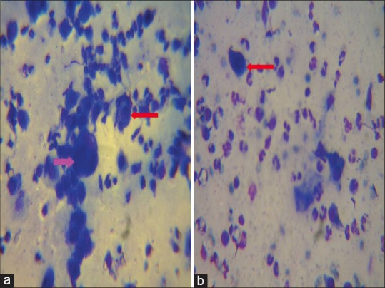
Figure 5.

Tzanck smear showing Multi Nucleated Giant cells (MNGs) (pink arrow) and Tzanck cells (red arrow) in herpetic infections (a) and Tzanck cells with numerous neutrophils in bullous impetigo (b). The stain used for preparing all the Tzanck smears was May - Grunwald-Giemsa stain (stock solution is prepared by diluting 1 part of stain with 3 parts of distilled water). Magnification used is 100X
