
Figure 6.
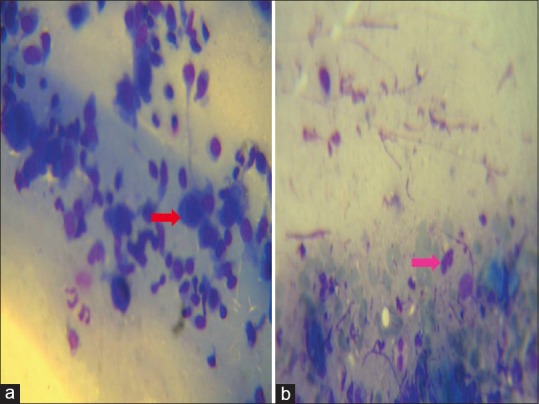
Tzanck smear showing Tzanck cells (red arrow) in pemphigus (a) and eosinophil (pink arrow) in bullous pemphigoid. The stain used for preparing all the Tzanck smears was May - Grunwald-Giemsa stain (stock solution is prepared by diluting 1 part of stain with 3 parts of distilled water). Magnification used is 100X