Figure 5.

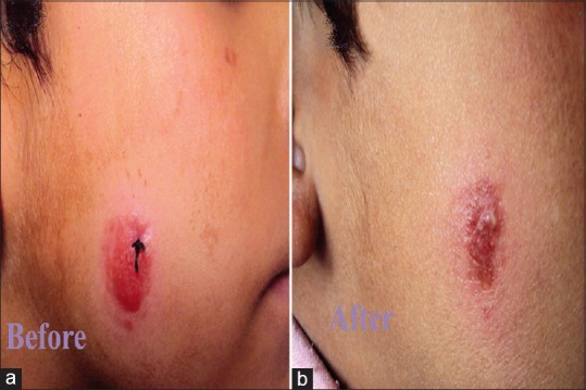
(a) Cutaneous leishmaniasis in a child showing nonulcerated nodular lesion. (b) Clearance of the lesion with atrophy and milia formation 3 weeks after treatment

(a) Cutaneous leishmaniasis in a child showing nonulcerated nodular lesion. (b) Clearance of the lesion with atrophy and milia formation 3 weeks after treatment