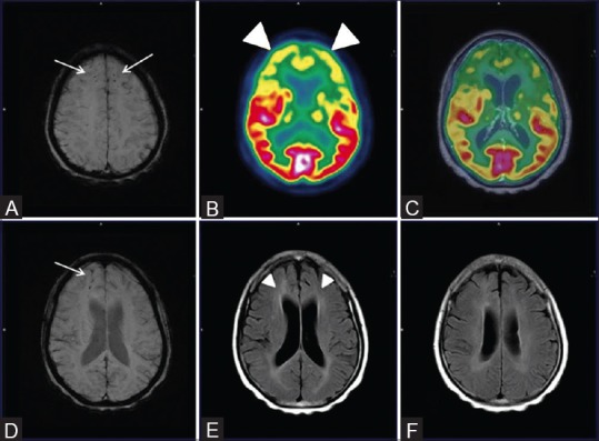
Figure 11 (A-F).

Axial SWI MRI Images (A and D) Reveal punctate old hemorrhagic residua in bilateral frontal and parietal regions (white arrows) with corresponding bilaterally symmetrical fronto-temporal hypometabolism (white arrowheads) on axial PET (B) and fused PET/MRI images (C) Also, on axial FLAIR MRI images (E and F) Ill defined non enhancing white matter changes in bilateral periventricular regions, corona radiata and centrum semiovale (white arrowheads) were also noted
