
Figure 1.
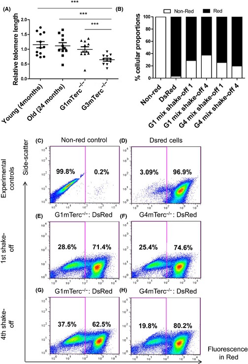
Telomerase ablation results in telomere shortening and a proliferative deficit in microglia. (A) Relative telomere length measurement in isolated microglia from young (4 months; n = 12) and old (24 months; n = 11) mice shows no significant difference, while isolated microglia from G1(n = 11) and G3 mTerc−/− (n = 11) mice (6 months) have significantly shorter telomeres in G3 mTerc−/− microglia. Asterisks * indicate comparisons for which P‐value was significant according to one‐way ANOVA *P < 0.05, **P < 0.005, ***P < 0.001 and ns, not significant. Error bars indicate standard deviation (SD). (B) G1 and G4 mTerc −/− pup brains (each group from six pooled pup brains) were used to prepare shake‐off glial cultures with 10 parts equivalent DsRed pup brain homogenates (pooled from 12 DsRed pup brains). Repopulation ability of G1 and G4 mTerc −/− microglia was assessed by quantifying the percentage of the red vs. nonred populations. Flow cytometry controls of nonred (C) and DsRed (D) cells. G4 mTerc −/− microglia showed a decreased % proportion on four successive shake‐off cultures (H vs. F), while G1 mTerc−/− increased in % proportion with respect to Ds Red (G vs. E) indicating compromised proliferation and repopulation capacity in G4 mTerc −/− microglia.