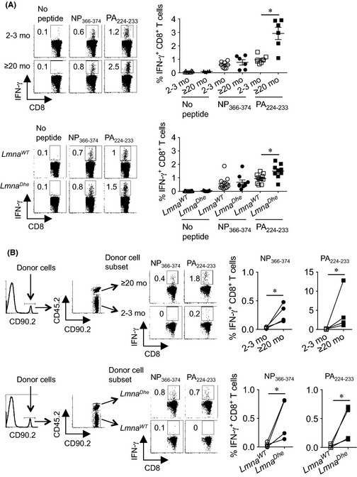
Figure 2.

Skewed immune dominance of influenza virus‐specific CD8+ T cells with aging is replicated in accelerated aging Lmna Dhe mice. (A) Representative FACS plots and composite analysis of percent IFN‐γ‐producing mediastinal lymph node CD8+ T cell after stimulation with each influenza‐specific MHC class I peptide compared with no stimulation controls day 8 after influenza A infection (3000 PFUs) in ≥20‐month compared with 2‐ to 3‐month‐old mice (top), or 2‐ to 3‐month‐old Lmna Dhe mice compared with littermate Lmna WT control mice (bottom). (B) Cell gating scheme, and composite analysis of percent IFN‐γ‐producing CD8+ splenocytes after stimulation with PA 224–233 or NP 366–374 peptides for ≥20‐month compared with 2‐ to 3‐month‐old donor mice (top), or 2‐ to 3‐month‐old Lmna Dhe mice compared with Lmna WT donor mice (bottom) day 8 after influenza A infection (3000 PFUs). These data are representative of results from at least two independent experiments with similar results. Bar, mean ± 1 SE. *, P < 0.05.
