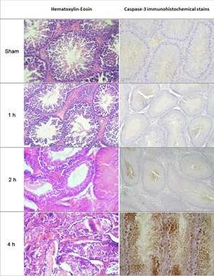
Figure 2.

Representative photographs of haematoxylin–eosin (H&E) (left column) and active caspase‐3 immunohistochemical (IHC) (right column) staining of four groups. H&E slides showed minor to serious damage at the seminiferous tubules, haemorragia at the interstitial area and loss of spermatogenic cells all increasing with time. Active caspase‐3 immunohistochemical staining clearly demonstrates apoptotic cells, stained as dark brown, increased to high numbers in some animals, especially at the 4‐h torsion group. (Magnification 240 X).
