Figure 1.

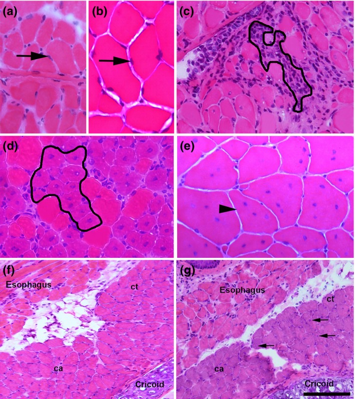
Histological appearance of a 9‐month‐old diaphragm (a) and quadriceps (b) muscle in controls, displaying fibres with peripheral nuclei (arrows). c, histological appearance of a 4‐month‐old mdx diaphragm with an area of inflammation outlined showing inflammatory cells. d, histological appearance of a 1‐month‐old mdx quadriceps with clusters of myofibres under regeneration (outlined), which are observed as plump cells with at least one centrally located nucleus and a small diameter; note the poor inflammatory infiltration. e, histological appearance of a 9‐month‐old mdx quadriceps showing centrally nucleated (arrowhead) polygonal cells with normal size. f and g, images from 18‐month‐old intrinsic laryngeal muscles in control (f) and in mdx (g). The posterior cricoarytenoid (ca) and cricothyroid (ct) muscles are shown. Some central nucleated fibres (arrows in g) in the non‐spared laryngeal muscle. Scale bar (shown only in g), 40 μm (a, b, c, e), 50 μm (d) and 210 μm (f, g). H&E‐stained representative sections.
