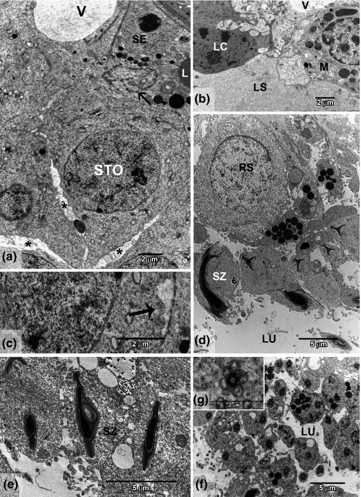
Figure 3.

Ultrastructure of testis intoxicated with cadmium (GCd). (a) Basal compartment showing altered structure with an irregular blood–testis barrier (thin arrow), lipid droplets (L) in Sertoli cell (SE) and spermatocyte (STO), vacuolated region (V) and intercellular spaces (*). (b) Interstitial region showing vacuole (V) in lymphatic space (LS), Leydig cell (LC) and macrophage (M). (c) Cytoplasmic degeneration (thick arrow). (d) Region of spermiation showing an immature spermatid (RS) and compact vesicles. (e) Vacuole in sperm head (SZ) with retained cytoplasm. (f) Lumen area (LU) showing retained cytoplasm around flagella and abundant compact vesicles. (g) Detail of round mitochondria surrounding the axoneme.
