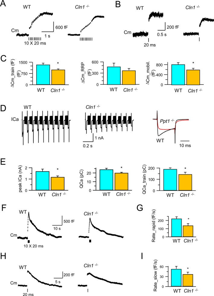
Figure 2.

Exocytosis, endocytosis and Ca2+ currents at the calyces of Held. Exocytosis of synaptic vesicles and RRP in the calyx of Held from both WT and Cln1 −/− mice were measured by monitoring the whole‐cell membrane capacitance (C m) (A and B). Compared to its WT littermate, Cln1 −/− mice had 25% reduction in exocytosis (upper panel), a small, but insignificant decrease in the RRP size (middle panel), and significant inhibition of vesicle mobilization of releasable vesicles (C). The depolarization‐evoked Ca2+ currents were also detected. (D) The peak amplitude (ICa) was reduced from 1.7 nA (WT) to 1.2 nA (Cln1 −/−); while the charge of Ca2+ influx was reduced from 24 pC (WT) to 20 pC (Cln1 −/−) for a single 20 msec pulse, and from 190 pC (WT) to 144 pC (Cln1 −/−) for a train of 10 pulses (E). Compared to the WT mice (F), Cln1 −/− littermates showed substantial reduction in rapid endocytosis (221 fF/sec vs. 136 fF/sec) (F and G) and in slow endocytosis (52 fF/sec vs. 37 fF/sec) (H and I). RRP, readily releasable vesicle pool; WT, wild‐type; ICa, calcium current. *means that the results are significantly different between wt and Ppt1 −/−, at the significance level: P < 0.05 (two‐tailed student t‐test).
