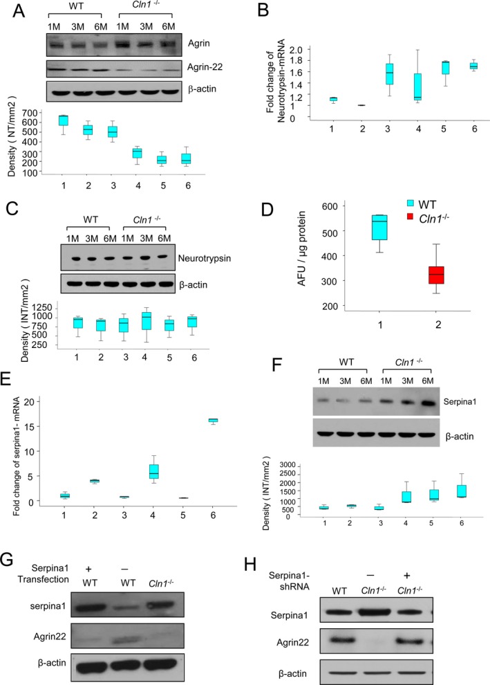
Figure 3.

Neurotrypsin‐mediated agrin‐22 production is disrupted in Cln1 −/− mouse brain. Western blots analysis of intact and cleaved agrin (agrin‐22) proteins in wild‐type (WT) and Cln1 −/− mouse brains (A). The lower panel showed that the mean densitometry of each agin‐22 from WT and Cln1 −/− mouse brains. Compared with WT mice the levels of agrin‐22 in the brain of Cln1 −/− littermates were markedly reduced (P < 0.05, Excel student t‐test) (A). Neurotrypsin‐mRNA levels measured by qRT‐PCR showed no significant difference between WT mice and their Cln1 −/− littermates (1 and 2 for 1‐month‐old WT and Cln1 −/− mice, respectively; 3 and 4 for 3‐month‐old WT and Cln1 −/− mice, respectively; 5 and 6 for 6‐month old WT and Cln1 −/− mice, respectively) (B). Western blot analysis showed that neurotrypsin protein levels between Cln1 −/− and their WT littermates were virtually no difference (C). Enzymatic activities of neurotrypsin in brain tissues of 6‐month old Cln1 −/− mice was significantly lower compared with those of their WT littermates (D). Quantitative RT‐PCR and Western blot analyses showed increased levels of serpina1‐mRNA (E; 1 and 2 for 1‐month‐old WT and Cln1 −/− mice; 3 and 4 for 3‐month‐old WT and Cln1 −/− mice; 5 and 6 for 6‐month‐old WT and Cln1 −/− mice, P < 0.05, Excel student t‐test) and protein (F) from cortical tissues of Cln1 −/− mice compared with those of their WT littermates. Serpina1 overexpression in WT mouse glial cells significantly reduced the level of agrin‐22 (G). To further confirm the inhibition of serpina1 to neurotrypsin activity, serpina1 knockdown in Cln1 −/− glial cells showed that agrin‐22 protein level was increased (H).
