
Figure 7.
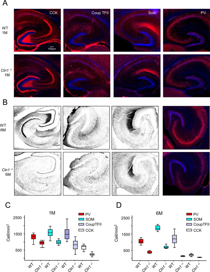
Loss of hippocampal interneurons during early development of Cln1 −/− mice. Representative images of hippocampal sections from WT mice (upper panels) and those from their Cln1 −/− littermates (lower panels) stained for GABAergic interneuron markers at 1 month (A and C) and 6 months of age (B and D). Markers examined were CCK, CoupTFII, SOM, and PV. Pooled group data for cell density counts of the labeled interneurons in WT and Cln1 −/− are provided in (C) and (D) for mice aged 1 and 6 months, respectively; (C) a total of 24 sections per marker from 3 different WT and Cln1 −/− mice were processed and counted parallel; (D) a total of 8–12 sections per marker from a WT mouse and those from a Cln1 −/− littermate were examined. Excel student t‐test was used for statistical analysis of the data, P < 0.01. WT, wild‐type; CCK, cholecystokinin; CoupTFII, COUP transcription factor 2; SOM, somatostatin; PV, parvalbumin.