Figure 6.

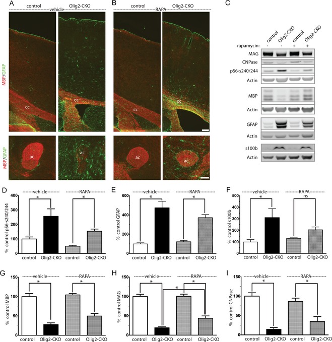
Improved myelination but persistent gliosis following rapamycin treatment. CKO and littermate control animals were treated with rapamycin 3 mg/kg, 5 days per week from P30 to P60. P60 sagittal brain sections were stained with MBP to identify myelin and GFAP to identify astroctyes (A, B) (cc – corpus callosum, ac – anterior commissure) Scale bar = 100 μm. Gliosis was increased as identified GFAP and s100β expression (C, E, and F) and remained significantly elevated despite rapamycin treatment (D). Expression of MAG improved with rapamycin treatment but did not completely normalize (C, G–I). Data represent mean ± SEM, n = 4 animals per group. *P < 0.05 by one‐way ANOVA followed by Tukey's multiple comparison test. CKO, conditional knockout; MBP, myelin basic protein; GFAP, glial fibrillary acidic protein; MAG, myelin‐associated glycoprotein; ANOVA, analysis of variance.
