
Figure 5.
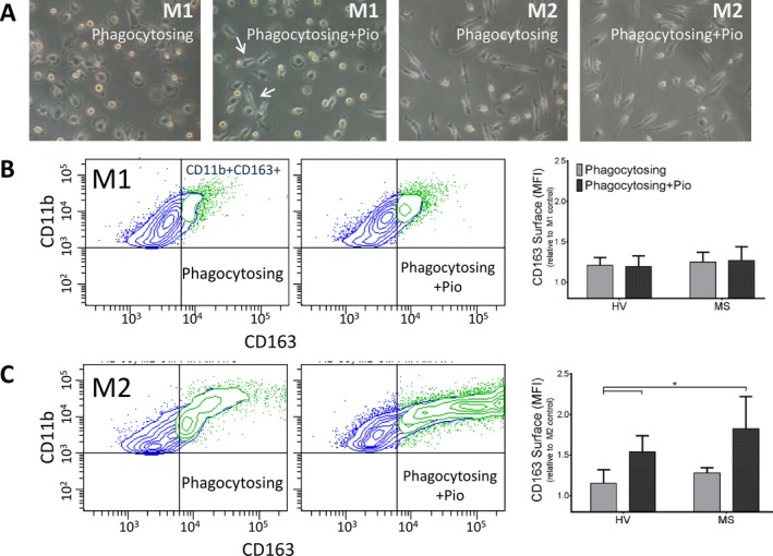
Pioglitazone treatment increases expression of the M2 surface marker CD163 on MS patient macrophages. (A) Light micrographs of M1 and M2 monocyte‐derived macrophages. M1 macrophages display a flattened, amoeboid phenotype with some cells elongated upon pioglitazone treatment (arrows). M2‐polarized macrophages display a bipolar appearance. (B) There is no change in CD163 expression in CD11b+ M1‐polarized, myelin‐phagocytosing macrophages compared to resting cells after treatment with 1 μmol/L pioglitazone. (C) M2, pioglitazone‐treated macrophages display increased CD163 expression upon myelin debris phagocytosis compared to HV macrophages, indicating further anti‐inflammatory polarization. Adjusted P‐values for pairwise comparisons in a adjusted P‐values for pairwise comparisons in a two‐way repeated measures ANOVA with Tukey's test. Mean ± SEM, *P < 0.05, n = 10/group. MS, multiple sclerosis; HV, healthy volunteer; ANOVA, analysis of variance.