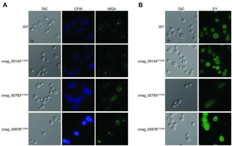
Figure 6. Cell wall chitin and chitosan exposure as assessed by staining of C. neoformans insertional mutants identified by AIM-Seq.
Strains were incubated in CO2- independent medium at 37°C for 18 hours prior to staining and imaging. Exposure of cell wall components was assessed by staining with (a) CFW and WGA for chitin, and (b) eosin Y for chitosan. Stained cells were imaged by fluorescent microscopy (630x).

