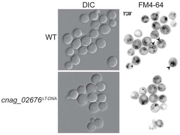
Figure 8. Vacuolar staining of WHM27/cnag_02676ΔT-DNA.
Strains were incubated in YPD liquid media at 30°C for 18 hours prior to staining and imaging. Cells were stained with the endocytosis marker, FM4-64, for 10 minutes on ice, followed by washing and incubation at room temperature for 30 minutes. Stained cells were imaged by fluorescent microscopy (630x). Black arrows indicate FM4-64 staining of vacuolar membranes.

