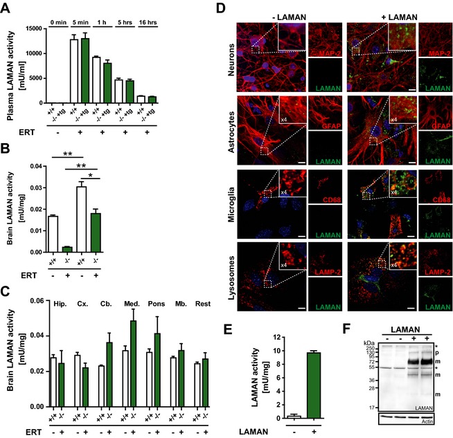
Figure 6.

Broad uptake of rhLAMAN into brain fully restores endogenous LAMAN activity. (A) Wild‐type (+/+) and transgenic knockout mice (‐/‐ tg+) received one single injection of 1000 U/kg and LAMAN plasma activity measured at different time (t) points (before injection [0], 5 min, 1 h, 5h, and 16 h). 16 h after injection mice were perfused with phosphate buffer to eliminate remaining enzyme from circulation and LAMAN activity determined in (B) whole brain and (C) isolated brain regions, respectively (*P < 0.05, **P < 0.01). (D) Mixed culture of neurons, astrocytes and microglial cells incubated with rhLAMAN (green) for 16 h. Neurons were visualized, using MAP‐2 as a marker (red), whereas astrocytes and microglia are highlighted by staining with markers against GFAP (red) and CD68 (red), respectively. Lysosomal localization of rhLAMAN is reflected by co‐staining with LAMP‐2 (red). (E) Enhanced LAMAN activity was observed in the same mixed cultures after treatment with the enzyme. (F) Similarly, uptake of the enzyme was verified via immunoblotting. (Scale bar: 10 μm). rhLAMAN, recombinant human lysosomal acid alpha‐mannosidase; LAMP‐2, lysosomal‐associated membrane protein 2.
