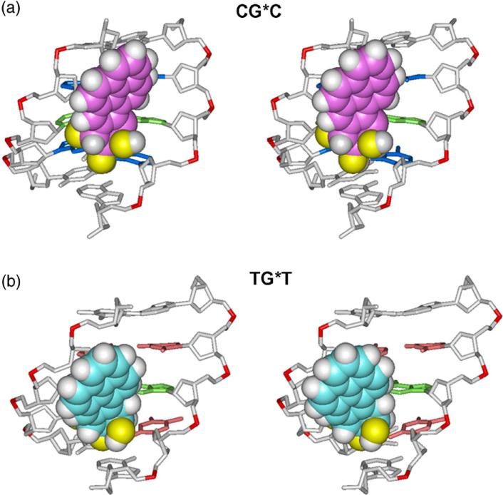
Fig. 2.
Stereo views of the 10S (+)-trans-anti-[BP]-N2-dG adduct looking into the minor groove in the (a) CG*C and (b) TG*T sequence contexts. The structures shown are the best representative conformations39 for the last 7.0 ns of the MD simulations. Only the central 5-mers are shown. In the CG*C sequence context, for the BP moiety, the carbon atoms are magenta, oxygen atoms yellow, and hydrogen atoms white. The modified guanine and its partner cytosine are green. The flanking base pairs are marine. In the TG*T sequence context, for the BP moiety, the carbon atoms are cyan, oxygen atoms yellow, and hydrogen atoms white. The modified guanine and its partner cytosine are green. The flanking base pairs are pink. The DNA duplexes are white, except for the phosphorus atoms, which are red. Hydrogen atoms and pendant phosphate oxygen atoms in the DNA duplexes are not displayed. The BP moieties are in CPK representation. The backbones of the duplexes were aligned to be optimally superimposed in order to highlight the different orientations of the BP rings.

