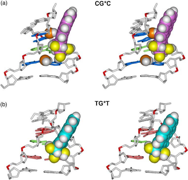
Fig. 3.
Effect of guanine amino groups in the minor groove on positioning of the 10S (+)-trans-anti-[BP]-N2-dG adduct in (a) CG*C and (b) TG*T sequence contexts. Only the central 5-mers are shown. The structures shown are the best representative conformations39 from the last 7.0 ns of the MD simulation. The BP moiety and relevant guanine amino groups are in CPK representation. Hydrogen atoms in the rest of the DNA duplexes are not displayed. The color scheme is the same as in Fig. 2, except that guanine amino groups are in orange (N) and white (H). Supplementary data movie_a and movie_b show these structures as they statically rotate, and movie_c and movie_d show their dynamics.

