Key Clinical Message
Nonobstetric hematomas of the vulva are rare and not extensively reported in literature. There are no consensus guidelines and a paucity of literature to guide best practices with regard to management. We present a case of vulva hematoma in a teenage girl. Our experience highlights the importance of prompt surgical intervention to reduce associated morbidity and minimize hospital stay.
Keywords: Conservative, evacuation, hematoma, trauma, vulva
Introduction
Vulvar hematomas are usually seen in the obstetric population following repair of episiotomies and birth‐related soft tissue injury. However, traumatic nonobstetric vulva hematomas are rare. They may arise secondary to blunt trauma sustained during a fall from height 1, 2, 3, sexual assault, foreign body insertion, and coitus 4, 5, 6, 7. Although most are small and pose little threat to the patient, nonobstetric hematomas may become large enough to cause hemodynamic instability. While conservative management is often the mainstay of treatment there is a subset of the population that will need surgical evacuation and repair. We detail the presentation and management of a young woman with a significant vulva hematoma following accidental blunt trauma.
Case Report
A healthy 16‐year‐old female, with no known comorbidities, presented to the Outpatient Department with severe perineal pain. The mechanism of injury was a fall from height in which the patient sustained blunt trauma to the perineum. The patient reported immediate onset of severe pain associated with a progressive swelling of the vulva and the presence of blood. This was associated with the gradual onset of headache and symptoms of presyncope, including generalized weakness, nausea, and lightheadedness. The patient denied any history of recent coitus or vaginal instrumentation. The patient was privately counseled on sexual assault, which she also denied. The patient was questioned regarding signs or symptoms suggestive of bleeding diathesis. The past medical history was noncontributory and the patient reported no known allergies or current medications. On physical examination the patient had an ashen appearance with pale sclera and mild respiratory distress. The heart rate was 115 bpm with a manual blood pressure of 100/60 mmHg and an oral temperature of 37.1°C. Her urine was clear with no evidence of macroscopic hematuria.
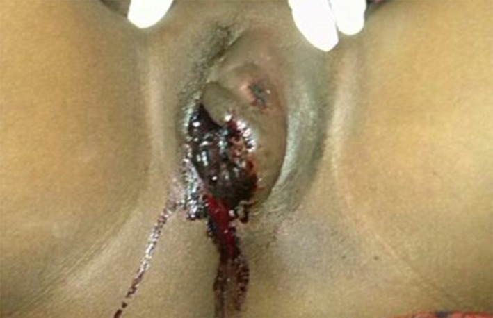
Examination of the perineum revealed a 10 × 8 cm swelling of the left vulva with right deviation of the right labia and clotted blood over the left labia minora (Fig. 1). There was no active bleeding per vagina. Basic laboratory investigations revealed a Hb of 10.2 g/dL, with a normal WBC and platelet count. The INR/PT was within normal parameters. Initial resuscitation consisted of 1500 mL IV bolus of crystalloid solution. Despite the infusion, the patient remained hypotensive and tachycardic. Given the degree of swelling and accompanying hemodynamic instability, the patient was consented for surgical intervention. Intraoperative, a large hematoma was evacuated through a medial incision of the left labia majora. A single, actively bleeding vessel was identified and suture ligated. The cavity was irrigated and inspected for hemostasis, which was subsequently closed with 1–0 chromic catgut (Fig. 2). Prior to the close of the case, a Foley catheter was inserted in the bladder. The patient had an uneventful postoperative course with prompt removal of urinary catheter and discharge on postoperative day three. The patient was seen in follow‐up at 4 weeks and the vulva appeared symmetric with the swelling completely resolved. The patient had no complaints.
Figure 1.

Preoperative: Swollen vulva with a clotted blood mass on left labia minora and lateral deviation of right labia majora.
Figure 2.

Postoperative: Mass evacuated with closure of dead space and an indwelling catheter.
Discussion
The vulva consist mostly of loose connective tissue and smooth muscle that is richly supplied by branches of the pudendal artery; a significant branch of the internal iliac artery 8. The venous drainage consists of labial veins, which are tributaries of the internal pudendal vein and venae comitantes. The injury to labial branches of the internal pudendal artery, which is located in the superficial fascia of the anterior and posterior pelvic triangle, can cause significant vulvar hematomas 9.
Blunt trauma to the vulva may occur secondary to reflex adduction of the thighs immediately preceding trauma. Its anatomical position and rich vascular supply make it susceptible to hematoma formation during perineal injury. In the case of a fall, a patient falls astride an object compressing the soft tissue of the vulva on the underlying pelvic girdle, which leads to laceration and hematoma formation. Sexual assault and forceful vaginal instrumentation are common causes of nonobstetric vulva hematoma. It is imperative to elicit the nature of the trauma in order to provide the most appropriate pre‐ and postoperative care. Social and cultural barriers to a thorough sexual history must be addressed to provide appropriate management.
The true incidence of nonobstetric vulvar hematoma is unknown but is likely underreported in the available literature. There are no consensus statements or best practice guidelines for the necessity or timing of surgical intervention. Propst et al. 10 observed that in the absence of acute hematoma expansion, nonoperative conservative management can yield good results. However, Benrubi et al. 11 found that conservative management of hematomas was associated with longer stays in hospital, an increased need for antibiotics and blood transfusion and greater subsequent operative intervention.
A hematoma that expands acutely is unlikely to settle with conservative management. Large vulva hematomas are best managed with surgical evacuation and primary closure 12. Kanai et al. 3 reported better outcomes with early surgical intervention. The hematoma should be evacuated and definitive hemostasis obtained 1, 5, 7. Selective arterial embolization is an appropriate alternative to traditional surgical exploration 2, 13. This may be particularly useful in the high‐risk operative candidate with prohibitive comorbidities, as the procedure does not require a general anesthetic. It may also be advantageous in the situation where the degree of soft tissue trauma makes careful delineation of the anatomy difficult 2, 14, 15, 16. Arterial embolization may also facilitate shorter hospital stays 17. However, this approach requires unique institutional resources that are often not available at the community or district level facility. Reported complications include groin hematoma, muscle pain, puncture site infection, guide wire perforation, and vagina fistula 16.
In the case presented, early surgical intervention is offered because of the enlarging hematoma and associated hemodynamic instability. For small, nonexpanding hematomas a conservative approach consisting of appropriate analgesia, cold compression, and close observation is appropriate. Prompt surgical intervention should be available if conservative management fails. Thus, transfer to a facility with surgical capability is essential 18. Surgical intervention is essential in the patient with a rapidly expanding hematoma or associated hemodynamic instability that does not immediately respond to crystalloid infusion.
Conclusion
In cases of severe nonobstetric blunt trauma to the perineum, hematoma of the vulva can represent a potentially life‐threatening hemorrhage that requires urgent surgical intervention. While conservative management plays an important role in minor perineal injuries, surgical hematoma evacuation and hemostasis is often desirable to minimize hospital stay and diminish the impact of long‐term complications.
Consent
Written informed consent was obtained from the patient for publication of this Case Report and any accompanying images. A copy of the written consent is available for review by the Editor of this journal on request. Permission to publish this case was also given by Iringa Regional Hospital.
Conflict of Interest
None declared.
Acknowledgment
The authors thank all the staff members of Department of Obstetrics and Gynecology for their support in preparation of the manuscript.
Clinical Case Reports 2015; 3(12): 975–978
References
- 1. Virgili, A. , Bianchi A., Mollica G., and Corazza M.. 2000. Serious hematoma of the vulva from a bicycle accident: a case report. J. Reprod. Med. 45:662. [PubMed] [Google Scholar]
- 2. Kunishima, K. , Takao H., Kato N., Inoh S., and Ohtomo K.. 2008. Transarterial embolization of a nonpuerperal traumatic vulvar hematoma. Radiat. Med. 26:168. [DOI] [PubMed] [Google Scholar]
- 3. Kanai, M. , Osada R., and Maruyama K. I.. 2001. Warning from Nagano: increase of vulvar hematoma and/or lacerated injury caused by snowboarding. J. Trauma 50:328. [DOI] [PubMed] [Google Scholar]
- 4. Habek, D. , and Kulas T.. 2007. Nonobstetrics vulvovaginal injuries: mechanism and outcome. Arch. Gynecol. Obstet. 275:93–97. [DOI] [PubMed] [Google Scholar]
- 5. Sau, A. K. , Dhar K. K., and Dhall G. I.. 1993. Nonobstetric lower genital tract trauma. Aust. N. Z. J. Obstet. Gynaecol. 33:433–435. [DOI] [PubMed] [Google Scholar]
- 6. Bechtel, K. , and Santucci K.. 2007. Walsh Hematoma of the labia majora in an adolescent girl. Pediatr. Emerg. Care 23:407–408. [DOI] [PubMed] [Google Scholar]
- 7. Dash, S. , Verghese J., Nizami D. J., Awasthi R. T., Jaishi S., and Sunil M.. 2006. Severe haematoma of the vulva–a report of two cases and a clinical review. Kathmandu Univ. Med. J. 4:228–231. [PubMed] [Google Scholar]
- 8. Palacios Jaraquemada, J. M. , Garcia Monaco R., Barbosa N. E., Ferle L., Iriarte H., and Conesa H. A.. 2007. Lower uterine blood supply: extrauterine anastomotic system and its application in surgical devascularization techniques. Acta Obstet. Gynecol. Scand. 86:228–234. [DOI] [PubMed] [Google Scholar]
- 9. Nelson, E. L. , Parker A. N., and Dudley D. J.. 2012. Spontaneous vulvar hematoma during pregnancy: a case report. J. Reprod. Med. 57:74–76. [PubMed] [Google Scholar]
- 10. Propst, A. M. , and Thorp J. M. Jr. 1998. Traumatic vulvar hematomas: conservative versus surgical management. South. Med. J. 91:144. [DOI] [PubMed] [Google Scholar]
- 11. Benrubi, G. , Neuman C., Nuss R. C., and Thompson R. J.. 1987. Vulvar and vaginal hematomas: a retrospective study of conservative versus operative management. South. Med. J. 80:991. [DOI] [PubMed] [Google Scholar]
- 12. Hwang, K. R. , Kim S. A, Kwon J. E, Jeon H. W., Choi J. E. and So Y. H.. 2014. A case of vulvar hematoma with rupture of pseudoaneurysm of pudendal artery. Obstet. Gynecol. Sci. 57:168–171. [DOI] [PMC free article] [PubMed] [Google Scholar]
- 13. Villela, J. , Garry D., Levine G., Glanz S., Figueroa R., and Maulik D.. 2001. Postpartum angiographic embolization for vulvovaginal hematoma: a report of two cases. J. Reprod. Med. 46:65–67. [PubMed] [Google Scholar]
- 14. Chen, T. H. , Chen C. H., Hong Y. C., and Chen M.. 2009. Peurperal pelvic hematoma successfully treated by primary transcatheter arterial embolization. Taiwan J. Obstet. Gynecol. 48:200–202. [DOI] [PubMed] [Google Scholar]
- 15. Egan, E. , Dundee P., and Lawrentschuk N.. 2009. Vulvar hematoma secondary to spontaneous rupture of the internal iliac artery: clinical review. Am. J. Obstet. Gynecol. 200:e17. [DOI] [PubMed] [Google Scholar]
- 16. Vegas, G. , Illescas T., Muños M., and Pérez‐Piñar A.. 2006. Selective pelvic arterial embolization in the management of obstetric hemorrhage. Eur. J. Obstet. Gynecol. Reprod. Biol. 127:68–72. [DOI] [PubMed] [Google Scholar]
- 17. Cheng, Y. Y. , Hwang J. I., Hung S. W., Tyan T. S., Yang M. S., Chou M. M., et al. 2003. Angiographic embolization for emergent and prophylactic management of obstetric hemorrhage: a four‐year experience. J. Chin. Med. Assoc. 66:727–734. [PubMed] [Google Scholar]
- 18. Lynch, T. H. , Martı′nez‐Piñeiro L., Plas E., Serafetinides E., Türkeri L., Santucci R. A., et al. 2005. EAU guidelines on urological trauma. Eur. Urol. 47:1–15. [DOI] [PubMed] [Google Scholar]


