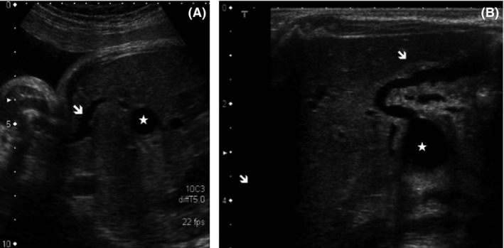
Figure 1.

(A): Ultrasound abdominal axial slice at 32 weeks of gestation, using a curved transducer. The cystic mass (white star) displays a thin wall in continuity with a small biliary canal and the gallbladder (white arrow). The wall of the gallbladder is irregular. (B): Abdominal axial slice performed at birth using with a high frequency linear probe. The same findings as those depicted prenatally are present [irregular and crenelated wall of the gallbladder (white arrow), cyst (white star)]. The intrahepatic biliary ducts are not dilated.
