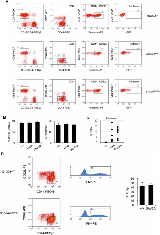
Figure 5.

Effector T cell differentiation in S100A‐deficient mice. A: Analysis of OVA‐specific T cell response in S100a4Gfp/Gfp, S100s4+/Gfp, or S100a4+/+ mice after infection with OVA‐trangenic L. monocytogenes. OVA‐H‐2Kb pentamer‐positive CD44+ CD62L− GFP+ CD8+ T cells were identified by flow cytometry. B: Relative numbers of OVA‐H‐2Kb pentamer‐positive CD44+ CD62L− effector CD8+ T cells isolated from S100a4Gfp/Gfp, S100s4+/Gfp, or S100a4+/+ mice after infection with OVA‐trangenic L. monocytogenes. Mean ± SD. n = 10 mice per group. C: Relative numbers of S100A4+ cells (% GFP+) within pentamer+ CD44+ CD62L− CD8+ effector T cells isolated from S100a4Gfp/Gfp, S100s4+/Gfp, or S100a4+/+ mice after infection with OVA‐trangenic L. monocytogenes. Mean ± SD. n = 10 mice per group. D: IFNγ production by CD44+ CD62L− CD8+ effector T cells isolated from S100a4Gfp/Gfp or S100a4+/+ mice after infection with OVA‐trangenic L. monocytogenes. T cells were isolated and restimulated in vitro with OVA for 16 h before flow cytometry analysis. Relative number of IFNγ is given (right panel). Mean ± S n = 10 mice per group.
