Figure 5.

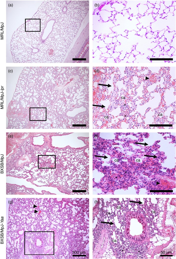
Histopathological features of the lungs in mice. Light microscopic photographs of haematoxylin & eosin (H&E)‐stained mouse lung sections of MRL/MpJ (a and b), MRL/MpJ‐lpr (c and d), BXSB/MpJ (e and f), and BXSB/MpJ‐Yaa (g and h), mice. The squares in (a), (c), (e) and (g) indicate the same areas as those in (b), (d), (f) and (h), respectively. A greater accumulation of mononuclear cells (asterisks), thicker interalveolar septa (arrows), congested blood vessels (arrowheads), and collapsed alveoli (ca) are visible in the lung tissue of MRL/MpJ‐lpr, BXSB/MpJ and BXSB/MpJ‐Yaa mice (c–h) than in that of MRL/MpJ mice (a and b). Bars = 300 μm (a, c, e and g). Bars = 50 μm (b, d, f and h).
