Figure 3.

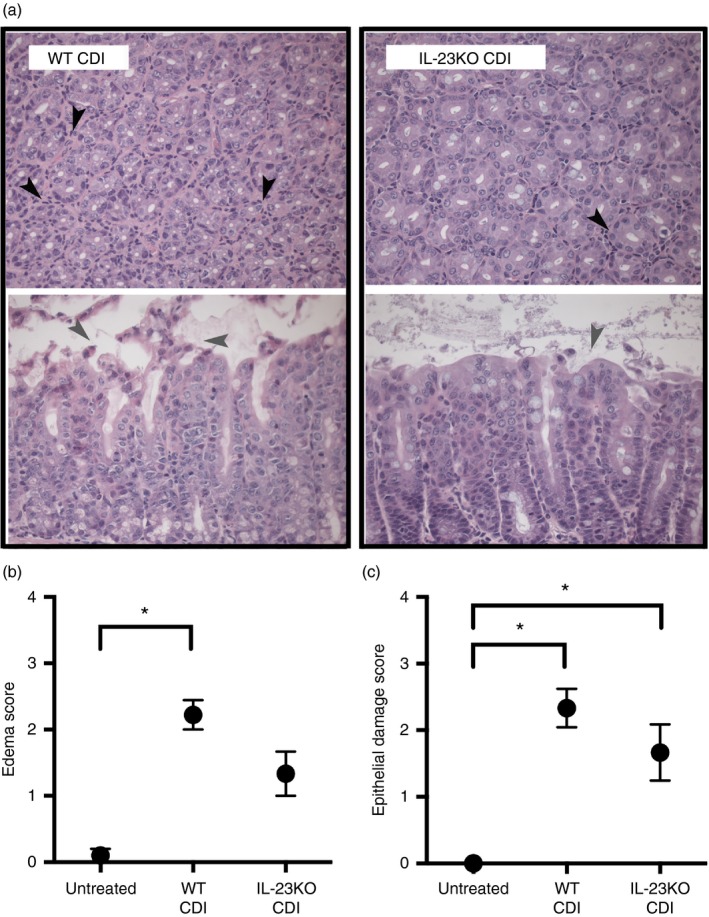
Colonic histopathology during Clostridium difficile infection in the absence of interleukin‐23 (IL‐23). (a) Representative photomicrographs of haematoxylin & eosin‐stained colonic sections from wild‐type (WT) C. difficile‐infected and IL‐23KO C. difficile‐infected animals. Cross‐sections of colonic crypts (upper images) and longitudinal sections of the epithelial–luminal interface (lower images) are shown for each genotype. Black arrowheads highlight cellular infiltrate, whereas grey arrowheads highlight epithelial damage. Total magnification for all images is 400×. (b, c) Histopathological scoring of colonic sections from Untreated, WT CDI, and IL‐23KO CDI mice. Slides were scored for oedema (b) and epithelial damage (c) as described in the Materials and methods. Data are shown as mean ± SEM. n ≥ 6 per group. CDI = C. difficile infected. Brackets indicate P < 0·05 for the differences between indicated groups.
