Figure 1.

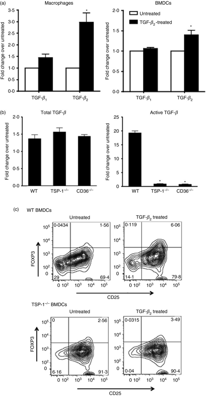
Transforming growth factor‐β 2 (TGF‐β 2) ‐exposed antigen‐presenting cells (APCs) predominantly express TGF‐β 2 and depend on thrombospondin‐1 (TSP‐1) to activate latent TGF‐β and induce Foxp3+ regulatory T (Treg) cells. (a) Expression of TGF‐β 1 and TGF‐β 2 in macrophages and bone marrow‐derived dendritic cells (BMDCs) cultured overnight in the presence of TGF‐β 2 as assessed by real‐time PCR. Changes in message levels relative to those in untreated APCs are presented. (b) Activation of TGF‐β by TGF‐β 2‐exposed wild‐type (WT), TSP‐1 null or CD36 knockout (KO) BMDCs detected using MFB‐F11 reporter fibroblast cells. Changes in the levels relative to the untreated APCs are presented. (c) Activation of CD4+ CD25− OT‐II T cells by ovalbumin (OVA) ‐pulsed WT or TSP‐1null untreated or TGF‐β 2‐treated BMDCs. Representative flow cytometry plots show percentage of activated Foxp3+ T cells (*P < 0·05 compared with TGF‐β 1 or total TGF‐β activated by WT cells). Data in panels a, b and c are as representative of three to four independent experiments.
