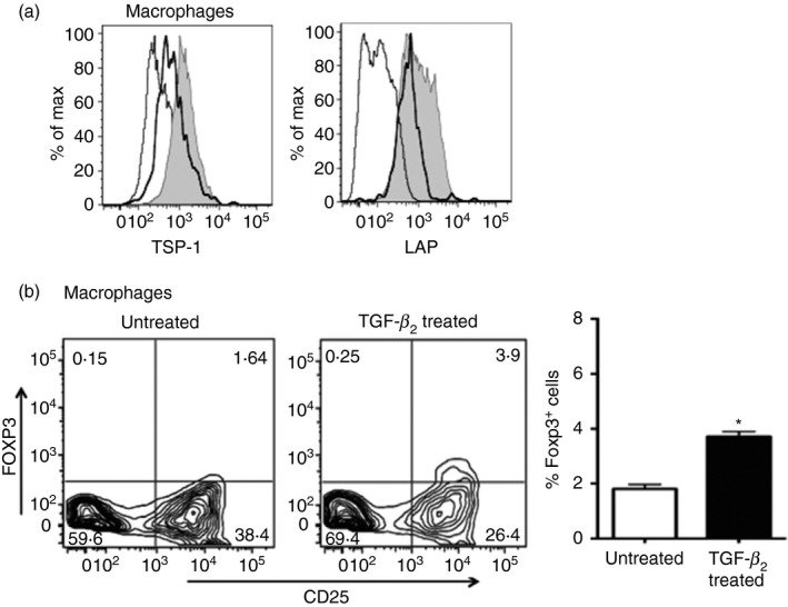
Figure 3.

The absence of thrombospondin‐1 (TSP‐1) receptor CD36 on transforming growth factor‐β 2 (TGF‐β 2‐exposed antigen‐presenting cells (APCs) reduces their ability to induce Foxp3. (a) Flow cytometric analysis of the surface staining of TSP‐1 and latency‐associated peptide (LAP) on macrophages derived from wild‐type (WT; filled grey) and CD36−/− (thick black lines) mice presented as histograms. Isotype control staining is indicated by thin lines. (b) Representative flow cytometric and mean data plots of Foxp3 induction by untreated or TGF‐β 2‐treated CD36−/− macrophages in CD4+ CD25− OT‐II T cells (*P < 0·05). Data are representative of three independent experiments.
