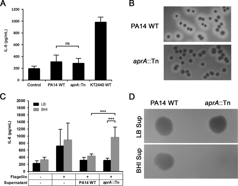
FIG 2.
Characterization of proteolytic activities of PA14 WT and aprA::Tn. (A) IL-8 response of A549-Gluc cells at an MOI of 10. The control represents A549-Gluc cells treated with PBS. (B) Qualitative analysis of protease activity using milk plate assays (LB agar supplemented with 1% skim milk). Zones of clearing surrounding colony growth indicate positive casein (a primary milk protein) hydrolysis. (C) Coincubation of A549-Gluc cells with PA14 WT flagellin pretreated with cell-free supernatant (Sup) samples obtained from cultures grown for 18 h in either LB or BHI. Data are presented as means from at least six experiments ± SD. Significance was evaluated by Wilcoxon test. ***, P < 0.001; ns, not significant. (D) Qualitative analysis of protease activity of cell-free supernatant samples obtained from cultures grown for 18 h in either LB or BHI, using skim milk assays (1% agarose gel supplemented with 1% skim milk).

