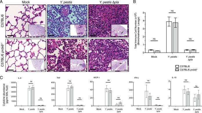
FIG 6.
Absence of Prdx6 does not alter the host immune response during Y. pestis respiratory infection. (A) Sections of formalin-fixed lungs stained with H&E from C57BL/6 or C57BL/6 prdx6−/− mice inoculated with PBS (mock), 104 CFU of Y. pestis, or 104 CFU of Y. pestis Δpla after 48 h. Representative images of inflammatory lesions are shown (arrows; n = 3). Bars, 100 μm (inset images) and 50 μm (larger images). (B) Enumeration of total immune cells present in the BALF of C57BL/6 or C57BL/6 prdx6−/− mice inoculated with PBS (mock), 104 CFU of Y. pestis, or 104 CFU of Y. pestis Δpla after 48 h. (C) Abundance of indicated inflammatory cytokines present in supernatants of the same BALF samples as those collected for panel B. Data are combined from 2 independent experiments (n = 10 for each group), and error bars represent the SEM (ns, not significant by one-way ANOVA with Bonferroni's multiple-comparison test).

