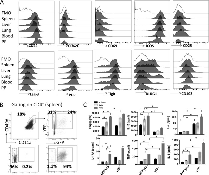
FIG 6.
CD4+ IFN-γ–YFP+ IL-10–GFP+ T cells exhibit a conserved phenotype in lymphoid and nonlymphoid organs but display tissue-dependent functional responses during malaria infection. IFN-γ and IL-10 dual-reporter mice were infected (i.v.) with 1 × 10⁴ P. yoelii NL pRBCs. (A) Representative histograms showing the expression of activation and regulation-associated molecules on CD4+ IFN-γ–YFP+ IL-10–GFP+ T cells in the different tissues on day 7 of infection. The results are the mean ± SEM for the group with 3 to 5 mice per group. The results are representative of those from 3 independent experiments. (B, C) Antigen-experienced (CD11a+ CD49d+) IFN-γ–YFP+ GFP+/− CD4+ T cells were sort purified from the spleens, liver, and lung of P. yoelii NL-infected (day 7, 1 × 10⁴ pRBCs [i.v.]) dual-reporter mice. (B) Representative dot plots showing the purity of sorted (splenic) CD4+ IFN-γ–YFP+ GFP− and CD4+ IFN-γ–YFP+ GFP+ T cells. (C) The sorted populations of tissue-derived cells were stimulated in vitro in triplicate with anti-CD3 and anti-CD28 for 24 h, and cytokine production in the culture supernatant was assessed using cytokine bead arrays. The results are the mean ± SEM for the group. The results are representative of those from 2 independent experiments. *, P < 0.05. Significance was tested using one-way ANOVA with Tukey post hoc analysis.

