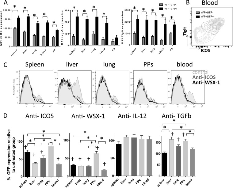
FIG 7.
IL-27R and ICOS control IL-10 expression by antigen-experienced CD4+ T cells in both lymphoid and nonlymphoid tissues during malaria infection. IFN-γ and IL-10 dual-reporter mice were infected (i.v.) with 1 × 10⁴ P. yoelii NL pRBCs. (A) The mean fluorescence intensity (MFI) of activation- and regulation-associated molecules on antigen-experienced CD4+ IFN-γ–YFP+ IL-10–GFP+ and CD4+ IFN-γ–YFP+ IL-10–GFP− T cells in the different tissues on day 7 of infection. (B) Representative dot plot showing the coordinated expression of Tigit and ICOS on splenic CD4+ IFN-γ–YFP+ IL-10–GFP+ T cells on day 7 of infection. (C, D) IFN-γ and IL-10 dual-reporter mice were treated with anti-ICOS, anti-WSX-1, anti-IL-12, or anti-TGF-β on days −1, 1, 3, and 5 of P. yoelii NL infection. (C) Representative histograms showing the expression of IL-10–GFP by parasite-specific CD4+ IFN-γ–YFP+ T cells in the different tissues on day 7 of infection following anti-ICOS or anti-WSX-1 MAb administration. (D) The frequencies of parasite-specific CD4+ T cells expressing IL-10–GFP in each tissue on day 7 of infection (relative to control treated group normalized to 100%). The results are the mean ± SEM for the group from 3 to 4 independent experiments (3 to 5 mice in each experiment). *, P < 0.05 compared with the control treated group; †, P < 0.05 compared with the control treated group. Significance was tested using an unpaired t test (A) and one-way ANOVA with Tukey post hoc analysis (D).

