FIG 8.

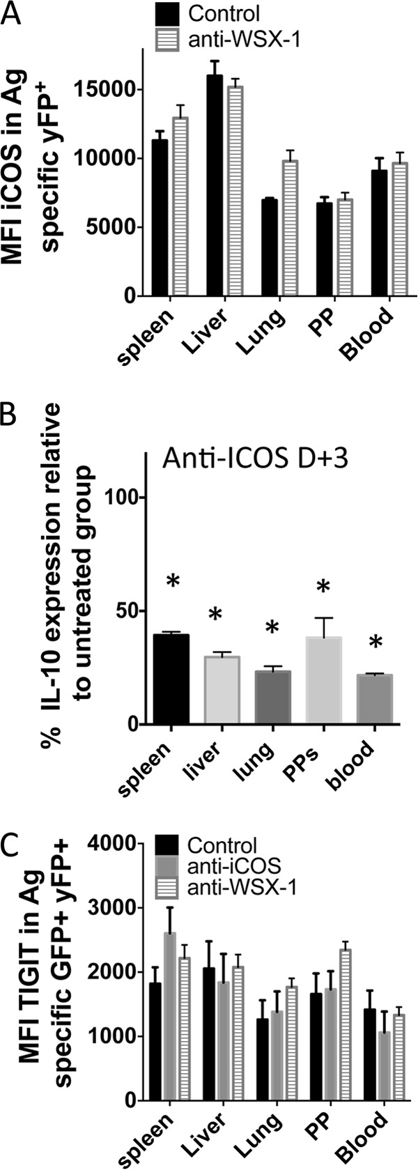
ICOS signaling after T cell priming actively promotes IL-10 expression by antigen-experienced CD4+ T cells during malaria infection. IFN-γ and IL-10 dual-reporter mice were infected (i.v.) with 1 × 10⁴ P. yoelii NL pRBCs. (A) The mean fluorescence intensity (MFI) of ICOS expression on parasite-specific CD4+ IFN-γ–YFP+ T cells in the different tissues of mice treated with control or anti-WSX-1 MAbs (MAb was administered on days −1, 1, 3, and 5 of infection, analysis was on day 7 of infection). (B) The relative frequencies of parasite-specific CD4+ T cells expressing IL-10–GFP in the tissues of mice treated with control or anti-ICOS MAbs (MAb was administered on day 3 [D+3] and day 5 of infection, analysis was on day 7 of infection). (C) The MFI of Tigit expression on parasite-specific CD4+ IFN-γ–YFP+ GFP+ T cells in the different tissues of mice treated with control, anti-WSX-1, or anti-ICOS MAbs (MAb was administered on days −1, 1, 3, and 5 of infection, analysis was on day 7 of infection). The results are the mean ± SEM for the group with 3 to 5 mice per group. The results are representative of those from 2 independent experiments. *, P < 0.05 compared with the control treated group. Significance was tested using an unpaired t test (A) and one-way ANOVA with Tukey post hoc analysis (B, C).
