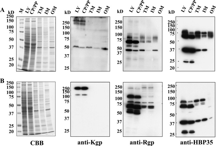
FIG 7.
Subcellular localization of CTD-containing proteins. P. gingivalis ATCC 33277 (A) and the Δomp17 strain (B) were subjected to fractionation, followed by SDS-PAGE and immunoblot analysis with anti-Kgp, anti-Rgp, and anti-HBP35 antibodies. M, marker; CP/PP, cytoplasm plus periplasm; TM, total membrane; IM, inner membrane; OM, outer membrane; CBB, Coomassie brilliant blue staining.

