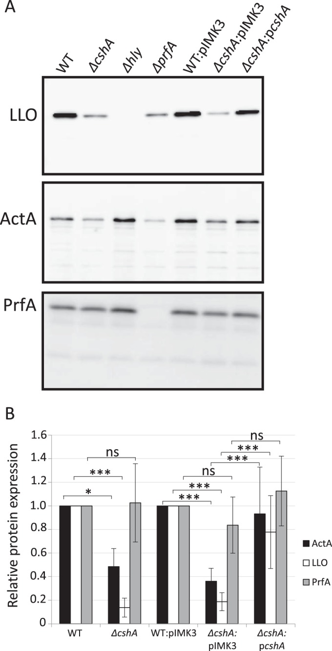
FIG 3.

Virulence factor expression in different strain backgrounds. (A) The indicated strains were grown at 37°C in pH-adjusted BHI (pH 5.5) to an OD600 of 0.9 before protein extraction, SDS-PAGE separation, and Western blot analysis. In the upper panel, the expression levels of LLO were analyzed using LLO-specific antibodies in samples of protein precipitated by trichloroacetic acid from filtered culture supernatants. In the middle panel, the expression levels of ActA were examined by extraction of boiled bacterial cells in Laemmli buffer using ActA-specific antibodies. In the lower panel, the PrfA level was examined from whole-cell fractions using PrfA-specific antibodies. (B) The expression of LLO, ActA, and PrfA, respectively, was quantified from panel A, and the results are shown relative to the wild type (WT) for EGDe or ΔcshA or to the WT::pIMK3 strain for WT::pIMK3, ΔcshA::pIMK3, or ΔcshA::pcshA, respectively. WT and WT::pIMK3 were arbitrarily set to 1.0. Error bars show the standard deviations. Statistics show two-tailed Student t test determinations (*, P < 0.05; ***, P < 0.001).
