
FIG 5.
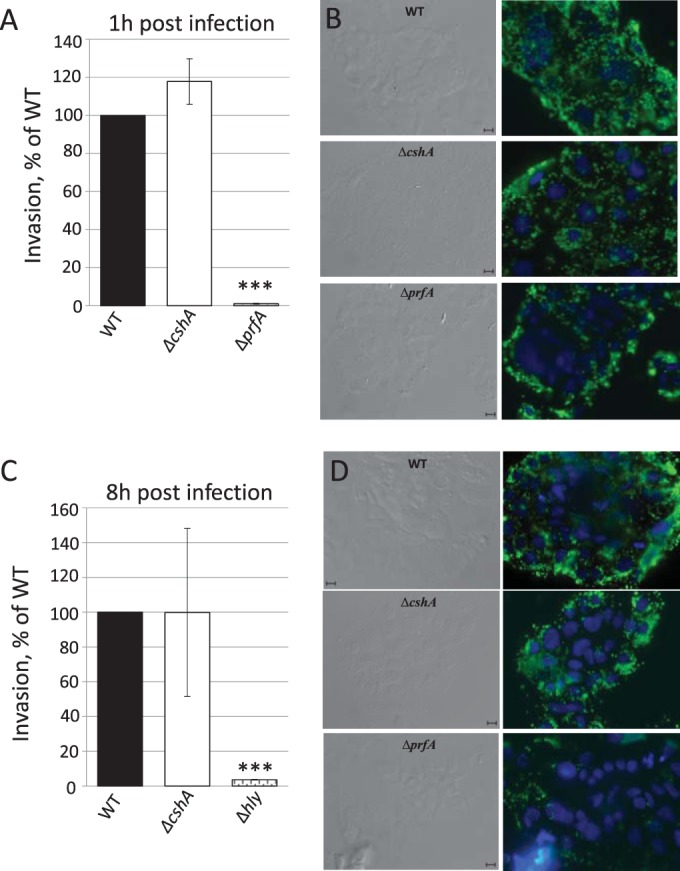
Infection assay. (A and C) Caco-2 cells were infected with wild-type, ΔprfA, Δhly, or ΔcshA strains for the indicated time points before cells were lysed, and bacteria were plated and counted. The infectivity of the ΔprfA, Δhly, and ΔcshA strains are shown relative to the wild-type strain (100%). Error bars show the standard deviations. All samples were compared to the wild-type using a two-tailed Student t test (***, P < 0.001). (B and D) Phase-contrast and fluorescence microscopy. The right panels show Caco-2 cells infected with the indicated strains for 1 h (B) or 8 h (D) and stained for Listeria (green) or cell nuclei (blue). The left panels show phase-contrast images of right panels. Bars, 10 μm.