FIG 5.
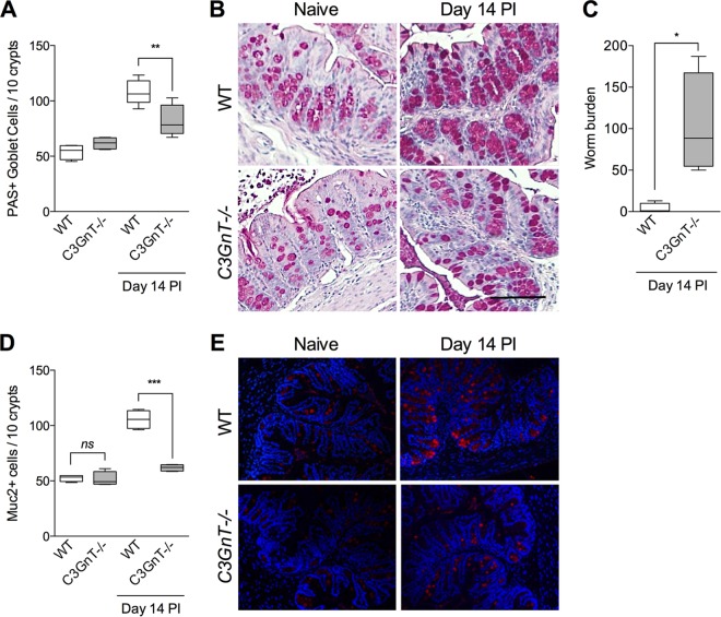
Numbers of PAS+ and Muc2+ cells were significantly lower in C3GnT−/− mice after T. muris infection. C3GnT−/− mice were infected with T. muris and sacrificed on day 14 p.i. (A) PAS+ goblet cell numbers in C3GnT−/− and wild-type mice on day 14 p.i. Groups were compared using Tukey's multiple comparison posttest. (B) Representative micrograph of PAS-stained goblet cells in colon sections. Magnification, ×200; bars, 100 μm. (C) Worm burden in C3GnT−/− and wild-type (WT) mice on day 14 p.i. The Mann-Whitney U test was used to analyze statistical differences among the groups. (D) Muc2+ cells as assessed by immunohistochemistry. (E) Representative images of Muc2-stained intestinal sections in C3GnT−/− and WT mice with or without T. muris infection. Groups were compared using Tukey's multiple comparison posttest. *, P < 0.05; **, P < 0.01; ***, P < 0.001; ns, not significant (5 mice per group).

