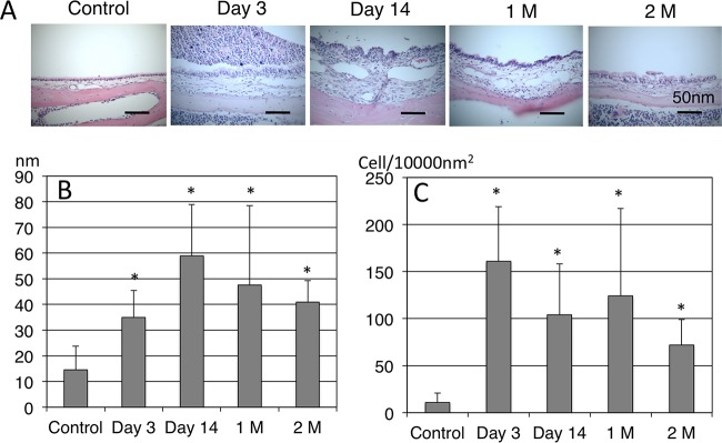
FIG 3.
(A) H&E image of the middle ear. In the control mice, few inflammatory cells were seen in the middle ear. At day 3 after inoculation, the MEM showed thickening and inflammatory cell infiltration containing polymorphonuclear cells, and these inflammatory responses were strong up to day 14. From 1 month (1M) to 2 months (2M) after inoculation, mucosal thickening and inflammatory cell infiltration continued with lymphocyte migration in the MEM. (B) Mucosal thickness of the MEM. (C) Number of inflammatory cells in the MEM. Compared to the control group, mucosal thickness of the middle ear increased significantly throughout the experiments. Inflammatory cell infiltration also increased throughout the experiments. *, P < 0.05 compared to the control by Mann-Whitney test.

