Figure 5.

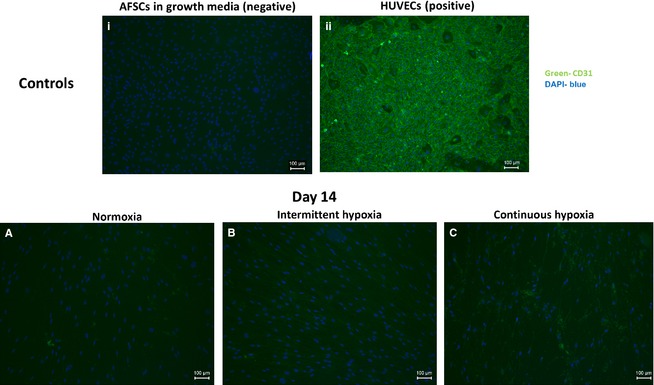
Fluorescent staining indicating CD31 expression by AFSCs in the presence of endothelial stimuli at day 14 of culture. No CD31 expression was visible in AFSC in growth media (i). Strong CD31 expression was visible in HUVECs (ii). AFSCs in normoxia (A), intermittent hypoxia (B) and continuous hypoxia (C) displayed weak expression of CD31 by day 14. AFSCs in growth media and HUVECs were used as the negative and positive controls, respectively. Images of AFSCs in growth media and of HUVECs were taken at day 14 of culture. Green represents CD31‐positive staining, blue represents DAPI nuclear stain. Scale bar: 100 μm.
