Figure 2.

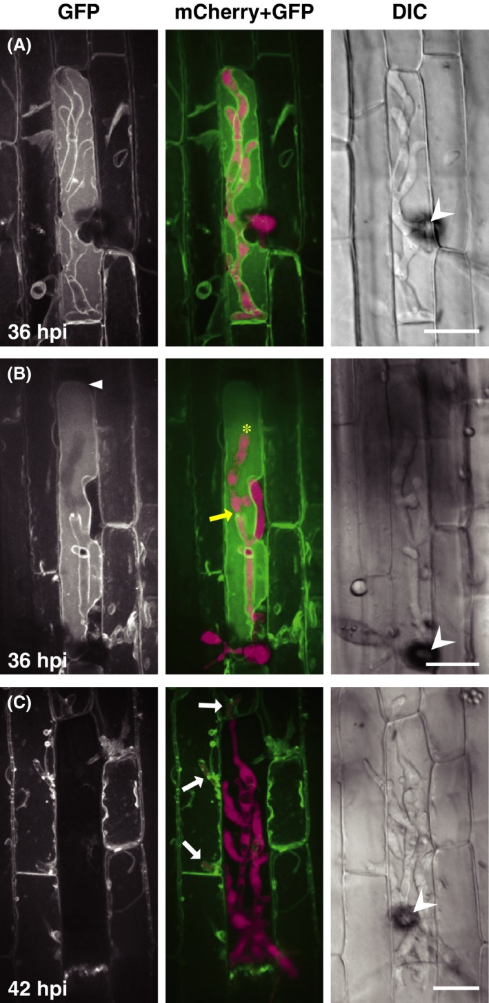
Invasive hyphae of Magnaporthe oryzae are surrounded by host vacuolar membranes. Leaf sheaths of transgenic rice plants constitutively expressing HvSUT2:AcGFP1 (vm‐GFP line) were inoculated with a conidial suspension of the compatible TmC line and observed using a laser confocal microscope. Invasive hyphae are outlined by vm‐GFP signals both in the primary‐ (A and B) and secondary‐invaded cells (C; white arrows). Vacuolar shrinkage is often observed (white arrowhead in B). Representative data of 5 (A), 6 (B), and 19 (C) similar images are shown. Asterisk and yellow arrow in the B (mCherry + GFP) panel indicate an invasive hypha inside the vacuole and the predicted point of penetrating across the vacuolar membrane, respectively. GFP, stacked z‐series confocal fluorescence images. mCherry + GFP, mergers of the GFP image, and stacked z‐series confocal fluorescence images of mCherry signals. Wedge, appressorium. Size bar = 20 μm. GFP, green fluorescent protein; DIC, differential interference contrast images.
