Figure 4.

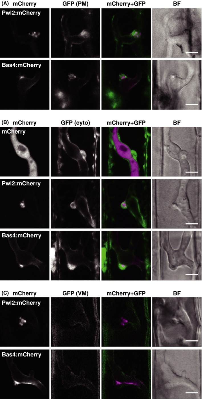
Confocal micrographs of the BIC. (A) Aggregation of the host plasma membrane‐derived at BIC. Leaf sheaths of transgenic rice plants constitutively expressing EGFP:LTi6b (pm‐GFP line) were inoculated with a conidial suspension of a compatible strain (Ina86‐137) transformed with PWL2p::PWL2:mCherry (PWL2mC line) or BAS4mC line. Representative data of 13 (PWL2mC) and 9 (BAS4mC) similar images are shown. (B) Accumulation of host cytosol at the BIC. Leaf sheaths of cyto‐GFP rice were inoculated with the compatible TmC, PWL2mC, or BAS4mC line. Representative data of 11 (TmC), 24 (PWL2mC), and 8 (BAS4mC) similar images are shown. (C) Surrounding of the BIC by host vacuolar membrane. Leaf sheaths of vm‐GFP rice were inoculated with the compatible PWL2mC or BAS4mC line. Representative data of 15 (PWL2mC) and 7 (BAS4mC) similar images are shown. These observations also reveal that Pwl2:mCherry is localized in BICs as puncta. All images are the single optical sections captured at 30 hpi. Size bar = 5 μm. BIC, biotrophic interfacial complex; GFP, green fluorescent protein; BF, bright field image.
