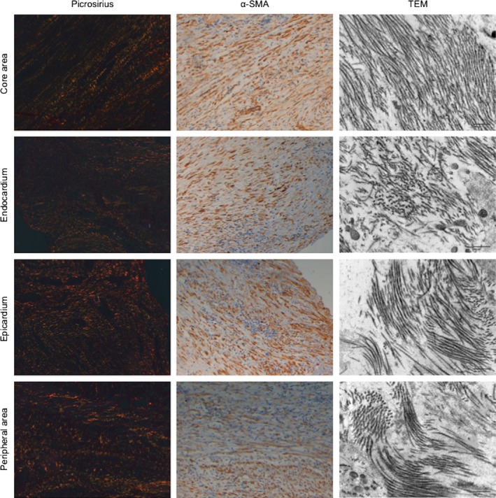
Figure 5.

Collagen fibers and myofibroblast arrangement in different areas of the fibrotic scar of swine samples. Left panels: Images stained with picrosirius photographed using a polarizer filter at 20× magnification. Middle panels: Images of alpha smooth muscle actin (α‐SMA) inmunohistochemistry photographed at 20× magnification. Right panels: Images of transmission electron microscopy (TEM) photographed at 10 000× magnification. Images exemplify the similarities between collagen and myofibroblasts arrangement in the different areas of the fibrotic scar. Upper panels correspond to the core area. In parallel with collagen fibers, myofibroblasts display a parallel disposition. TEM captions confirm the organized disposition of collagen fibers in the core area. Nevertheless, in the endocardium, epicardium and peripheral areas, collagen fibers stained by picrosirius display a disorganized disposition and myofibroblasts mirror this disorder. Finally, by TEM the collagen fibers are disposed in a disorganized manner. α‐SMA, alpha smooth muscle actin; TEM, transmission electron microscopy.
