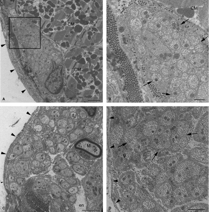
Figure 9.

Electron micrographs of intrinsic nerves in rabbit ventricles. (A) A profile of a small epicardial nerve; (B) the enlarged image of the boxed area (A). Note the oblong profile of the nerve with densely packed axons, which is located beneath the mesothelial cell layer (arrows) and is sheathed by dense collagen fibres with fibroblast processes instead of perineurium. Arrowheads in (B) point to groups of axons cuddled together in a polyaxonal pocket of Schwann cell. (C) A thick fibroblast‐ensheathed myocardial nerve from the septomarginal trabecula. Note the singular myelinated NF (M). (D) A profile of an endocardial nerve ensheathed by fibroblasts (arrowheads) with unmyelinated NFs that cuddle axons together in a polyaxonal pocket (arrows). Other abbreviations: CM, profiles of cardiomyocytes; en, endoneurium. Scale bars: 2 μm (A and C); 1 μm (D); 500 nm (B).
