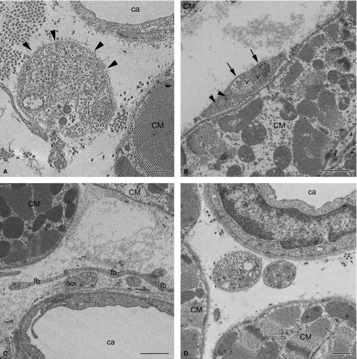
Figure 11.

Electron micrographs of myocardial NFs in the rabbit ventricles. (A) Unmyelinated NF with 12 axons (labelled with Arabic numerals) in contact with each other and enclosed by a Schwann cell from just one side (arrowheads) near a capillary (ca) and a cardiomyocyte (CM). Note that one of the axons contains neurotransmitter vesicles (V). (B) Unmyelinated NF with few axons near a cardiomyocyte (CM). Note that two axons with transmitter vesicles (V) are separated from a cardiomyocyte by a Schwann cell, but are open (arrows) to the more distant myocyte (CM) seen in the upper left corner of the panel. Some axons are significantly smaller in diameter than others, and are in contact with each other (arrowheads). (C) Two singular axons near a capillary (ca) are separated from a cardiomyocyte (CM) by fibroblast processes (fb). One axon (ax) is only in part closed by a Schwann cell (Sch), the other one (ax) is completely unenveloped. (D) Terminal axon (ax) filled with neurotransmitter vesicles without any Schwann cells close to cardiomyocytes (CM). Scale bar: 1 μm (A–C); 500 nm (D).
